Introduction:
Osteochondroma is a benign bone tumor. Benign means that it is not cancer. Benign tumors are collections of abnormal cells that stay in one place and do not move to other parts of the body. Osteochondromas form on the surface of the bone near the growing end (growth plate). They may feel like hard bumps near joints. An osteochondroma is made of bone and cartilage from the growth plate. The bump gets bigger as your child grows. Once your child stops growing, the osteochondroma stops growing too.
Many osteochondromas can be treated without surgery. A solitary (only one in the body) osteochondroma can be removed if it causes pain or other problems.
Some patients have many osteochondromas all over the body. This is called multiple osteochondromatosis. It is also known as multiple hereditary exostoses (MHE) or familial osteochondromatosis. MHE is caused by a problem in the EXT gene. Genes get passed to family members (inherited) so there are often other family members with MHE.
Description:
Osteochondromas are made of both bone and cartilage. Cartilage is the smooth lining of your joints that allows them to move easily. Osteochondromas occur near the ends of growing bones. They are usually found in the arms or legs, but they can form on any bone.
Osteochondromas are probably caused by a leftover piece of the growth plate that grows in the wrong direction. Osteochondromas can be flat (sessile) or have a long stalk (pedunculated).
Osteochondromas are often first noticed when the parent or patient feels a bony lump. There is usually no pain but sometimes it hurts when tendons and muscles slide over the bump. Some osteochondromas are found incidentally on X-rays when someone is being evaluated for an injury or pain.
Examination:
Your doctor will talk to you and your child about his or her medical history and will want to know if the bumps bother your child. The doctor will ask about any pain with activity or at night.
Your doctor will see if there are any bony lumps and feel them to check if they are painful. He or she will also check the motion in the joints near the osteochondromas.
Studies:
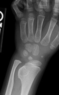
Figure 1 – X-ray showing an osteochondroma in the wrist.
A CT scan or MRI (Figure 4) may be ordered. This is helpful if the osteochondroma is in an area that is difficult to reach with surgery.
Treatment:
The treatment of solitary osteochondromas compared to MHE is very different, so these will be discussed separately.
SOLITARY OSTEOCHONDROMAS
Nonsurgical
If a solitary osteochondroma is not causing any problems, it does not need to be removed. Your doctor may want to see your child back and repeat X-rays to make sure the bump is not growing too quickly and causing problems (Figures 2 & 3). If the osteochondroma is causing pain or loss of motion, your doctor may recommend surgery.
A normal osteochondroma will not grow after your child stops growing. Children need to be taught that if their bumps get bigger once they become adults or if they start having pain, they need to see a doctor. It is rare for osteochondromas to become cancer in adults, but pain and tumor growth are the first signs of the development of cancer in adulthood.
Surgical
Osteochondromas that bother a child can be removed with surgery. While your child is asleep in the operating room, your surgeon will make an incision over the osteochondroma and remove it from the bone (Figure 5).
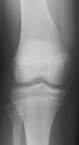
Figure 2: X-ray of a 12 year old female with pain during basketball. An arrow points to an osteochondroma located near the femur’s growth plate.
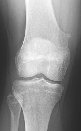
Figure 3: X-ray of patient in Figure 2 with continued pain now at age 15 years. The osteochondroma has changed over time.
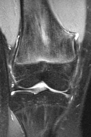
Figure 4: MRI of knee seen in Figure 3. MRI was obtained to evaluate internal structures of knee. Bursa and prominence adjacent to muscle are seen.
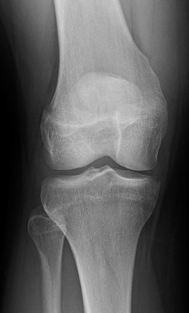
Figure 5: X-ray of knee of patient in Figures 2-4 after surgical removal of osteochondroma.
MULTIPLE HEREDITARY EXOSTOSIS (MHE)
MHE causes many of the bones in the body to grow abnormally. Patients with MHE are often shorter than average and may have one leg or arm longer than the other. MHE around the joint can also cause the bones to grow crooked (Figure 6). Osteochondromas in MHE are at higher risk of becoming cancer than solitary osteochondromas.
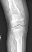
Figure 6 – X-ray of knee of a child with MHE. The knee is crooked, and there are multiple osteochondromas near the growth plates in all of the bones surrounding the knee.
Nonsurgical
Osteochondromas that are not causing problems do not need to be removed. However, if any osteochondroma is causing pain or getting much bigger, your doctor may recommend surgical removal.
Surgical
While your child is asleep in the operating room, your surgeon will make an incision over the osteochondroma and remove it from the bone – the same procedure as with a solitary osteochondroma.
If the bones are growing crooked, your doctor may recommend surgery to straighten the bones. This may consist of cutting the bone to re-align it or even lengthening or shortening a bone.
More Information
Condition QR Code:


